Legend : Nucleolar Structure
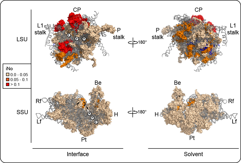
3-D models of human ribosomal subunits based on PDB entries 3J3D, 3J3A, 3J3F, and 3J3B. R-proteins are color-coded according to their iNo values. Left, subunit interface views; right, solvent exposed views. The aminoacyl (A), peptidyl (P) and exit (E) tRNA sites are indicated. Morphological features of the subunits are highlighted. On the LSU: the L1-stalk, the central protuberance (CP) and the phospho-stalk (P-stalk). On the SSU, the beak (Be), head (H), platform (Pt), body (Bd), lef foot (Lf) and right foot (Rf).
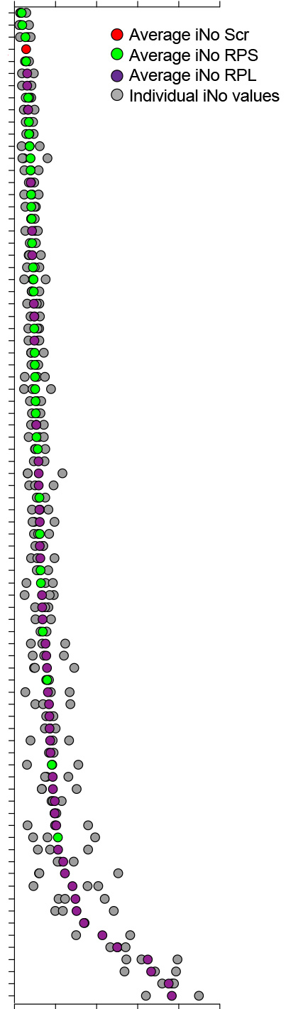
The histogram shows r-proteins classified according to their index of nucleolar disruption, iNo.
Colored-dots are mean of 3 individual experiments (shown in grey).